
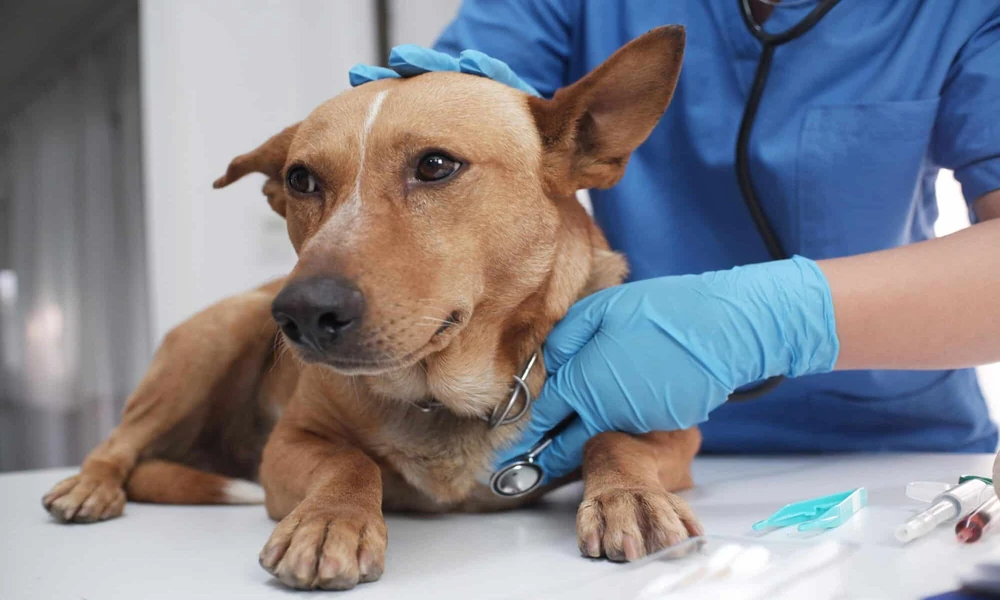
You know something is wrong, but the regular X rays did not give you a clear answer. Your dog is still limping, your cat still seems sore, and you are left watching for tiny signs that something hurts. That kind of waiting wears on you. If you are looking for a veterinarian in Cary, NC, you want proof, not guesses, and you want a plan that makes sense before your pet gets worse.
Bone scintigraphy for pets is often the next step when standard radiographs do not explain ongoing pain, lameness, or a suspected bone problem. It is a nuclear imaging test that can show changes in bone activity before those changes become obvious on X rays. In plain terms, it helps veterinarians look past the limits of a flat image and find trouble that may be hiding in plain sight.
Bone scintigraphy shows what standard X rays can miss
X rays are useful, fast, and common, but they only show structure. They are best at showing changes once enough damage, healing, or bone loss has already happened. That gap matters when your pet has early stress injury, subtle inflammation, infection, or pain that is real but not yet visible on a standard image.
Pet bone scans work differently. A small amount of radioactive tracer is injected, and that tracer collects in areas where bone is more active. A special camera then detects those hotspots. If your pet has a tiny fracture, bone infection, joint overload, or a hidden source of pain, the scan may light up that area even when an X ray still looks normal.
This is often where frustration turns into clarity. A dog that has been lame for weeks may have normal X rays and a normal orthopedic exam in the exam room, yet a bone scan can reveal a shoulder injury, a spinal issue, or a stress reaction in a limb. A cat that seems withdrawn and reluctant to jump may have a painful area that is hard to pinpoint without more sensitive imaging.
Veterinary teaching hospitals and specialty centers often use advanced imaging to sort out these cases. You can learn more about hospital imaging services through Cornell University’s imaging service, the University of Florida diagnostic imaging service, and Auburn’s radiology service.
Hidden bone pain in pets often carries a cost beyond the diagnosis
When a diagnosis drags on, the stress is not only medical. It is emotional and financial too. You may already have paid for repeat exams, pain medication, rest periods, and follow up X rays. You may be trying to keep an active dog calm for weeks without knowing whether rest is helping or just delaying the real answer.
That uncertainty can lead to harder choices later. A working dog with an undetected stress injury may keep compensating until a second limb is strained. An older pet with suspected bone infection or cancer may lose valuable time if the problem is not identified early. Even when the final answer is not simple, a better map of where the pain is coming from helps your veterinarian choose the right next test or treatment.
Veterinary bone imaging is not the first test for every pet, and it should not be. It is most useful when the problem is still unclear after an exam and standard imaging, or when there are several possible painful sites and your veterinarian needs to narrow the search.
Practical differences between standard X rays and bone scintigraphy
| Feature | Standard X Rays | Bone Scintigraphy |
|---|---|---|
| What it shows best | Bone structure, obvious fractures, advanced arthritis, clear masses | Areas of increased bone activity, early injury, hidden inflammation, multifocal pain |
| When it helps most | First line imaging for many orthopedic problems | When signs continue but X rays are normal or unclear |
| Sensitivity for early change | Lower | Higher |
| Specificity | Can be specific if the lesion is clear | May show where the problem is, but other tests may still be needed to confirm the cause |
| Typical use | Clinic and hospital settings | Usually specialty or referral hospitals |
| Cost and logistics | Usually lower cost and easier to access | Usually higher cost, more planning, and special handling after tracer use |
The tradeoff is straightforward. Bone scintigraphy is more sensitive, but it is not always the final answer by itself. A hotspot can tell your veterinary team where something is active, yet that area may still need CT, MRI, ultrasound, lab work, or biopsy to explain exactly why.
Bone scans for animals fit best in specific cases
This test tends to be most helpful in pets with persistent lameness, poor performance, back pain, suspected stress fractures, possible bone infection, or cases where pain seems to shift or involve more than one site. It can also help screen the whole skeleton when the source of pain is not obvious.
The phrase bone scan for animals can sound intimidating, especially because it involves nuclear medicine. For most owners, the practical concern is simpler. You want to know whether the test is worth the effort. That depends on whether the result will change treatment. If the answer could shift your pet from more rest to surgery, from broad pain control to targeted rehab, or from uncertainty to a specific diagnosis, the test may be worth serious discussion.
Three steps to take before deciding on advanced imaging
1. Ask what question the scan is meant to answer. A good reason for bone scintigraphy is not “we are out of ideas.” A good reason is “we need to locate the source of pain that standard imaging did not show” or “we need to see whether more than one area is involved.”
2. Review what has already been done. Gather prior X rays, exam notes, blood work, and videos of your pet walking at home. Small details matter. A specialist can compare old findings with current signs and may spot a pattern that changes the plan.
3. Talk through the full path, not just the test. Ask about sedation or anesthesia, expected cost, isolation rules after the tracer, and what follow up tests might come next. A scan is most useful when you understand what decisions it will help make.
Clear answers can change the course of care
When your pet is hurting and basic imaging has not explained why, it is hard to stay patient. You are not overreacting by wanting a deeper look. Bone scintigraphy gives your veterinary team another way to find pain that standard X rays can miss, and that can mean earlier treatment, fewer false starts, and a more direct path forward.
If your pet still has unexplained lameness or bone pain, contact your animal hospital and ask whether advanced imaging or referral for bone scintigraphy makes sense for the case.

Comments are closed.